On this page
Advice to improve autism by increasing synaptic pruning skips the most important question: does that person need more pruning at all? Researchers haven't established a single pruning pattern across autism, and a laboratory finding can't tell a family which connections a child's brain should keep or remove.
Synaptic pruning and autism are connected through a developing research hypothesis, not a clinical instruction. Understanding what scientists counted, which cells they studied, and what their models leave out makes it easier to separate interesting biology from promises that go beyond the evidence.
What the Main Studies Actually Measured
Autism isn't a settled case of too many synapses caused by too little pruning. Some human tissue findings support reduced elimination of connections, but they don't establish a universal cause or a way to diagnose an individual.
Synaptic pruning means the developmental removal of some connections between nerve cells. Calling it cleanup is useful shorthand, provided we don't assume that every removed connection was useless or that more removal is necessarily better.
A widely discussed 2014 postmortem study, described by Columbia University Irving Medical Center, found that dendritic spine density declined by approximately 16% in the autism group versus 45% in the comparison group from childhood to adolescence. Those are group-level differences across donated tissue samples, not measurements taken repeatedly from the same living children.
Synaptic pruning findings and what they do not prove
| Evidence | What was measured | Pruning direction | What it does not prove |
|---|---|---|---|
| Postmortem cortex from childhood through adolescence | Age-related differences in dendritic spine density | Less elimination in that sample | That weak pruning causes autism, or every autistic brain has extra synapses |
| Adolescent tissue from the same study | Lower LC3-II and persistently elevated mTOR-related signaling | Cellular recycling may be less efficient | A blood, scan, or clinic test of a person's pruning status |
| SCN2A-deficient mice and human organoids | Greater removal of postsynaptic material, with complement C3 implicated in mice | More elimination in that genetic context | A universal over-pruning subtype or a treatment that should reduce pruning |
| ADNP microglia-neuron screen | Changes in microglial uptake and synaptic pruning | A gene can modify pruning, without establishing a universal direction | That the uptake mechanism is fully mapped or ADNP status equals an autism test |
| Living people in clinic | No validated clinical assay of brain-pruning status | Direction cannot be assigned clinically | A basis for confirming or ruling out autism through synapse counts |
A spine count is an indirect measure
Researchers counted dendritic spines, small structures that generally receive excitatory signals. These counts provide a useful estimate of a particular kind of synaptic connection, not a complete inventory of every connection in the brain.
The human tissue sample was small and region-specific. Finding more remaining spines in selected cortex doesn't establish that the same pattern occurs throughout the brain, in every autistic person, or throughout life.
The molecular measurements added a possible explanation. LC3-II is associated with cellular recycling, while mTOR participates in growth-related signaling; the pattern was consistent with less efficient removal rather than necessarily excessive initial formation.
But consistency with a mechanism isn't proof of causation. Researchers couldn't determine from this tissue alone whether altered recycling contributed to autistic development, followed another developmental difference, or reflected several interacting processes.
The working rule is narrow but useful: pruning differences are biologically plausible in subsets of autistic people, not a biomarker or proof of a universally overconnected brain.
How the Brain Trims Connections in Childhood
Think of a gardener thinning a crowded hedge. The aim isn't to remove the most branches possible, but to preserve a workable arrangement while growth continues.
That analogy helps explain pruning, but it has limits. A brain doesn't have a gardener who knows in advance which connection a person will need, and autistic development shouldn't be understood as a badly maintained garden.
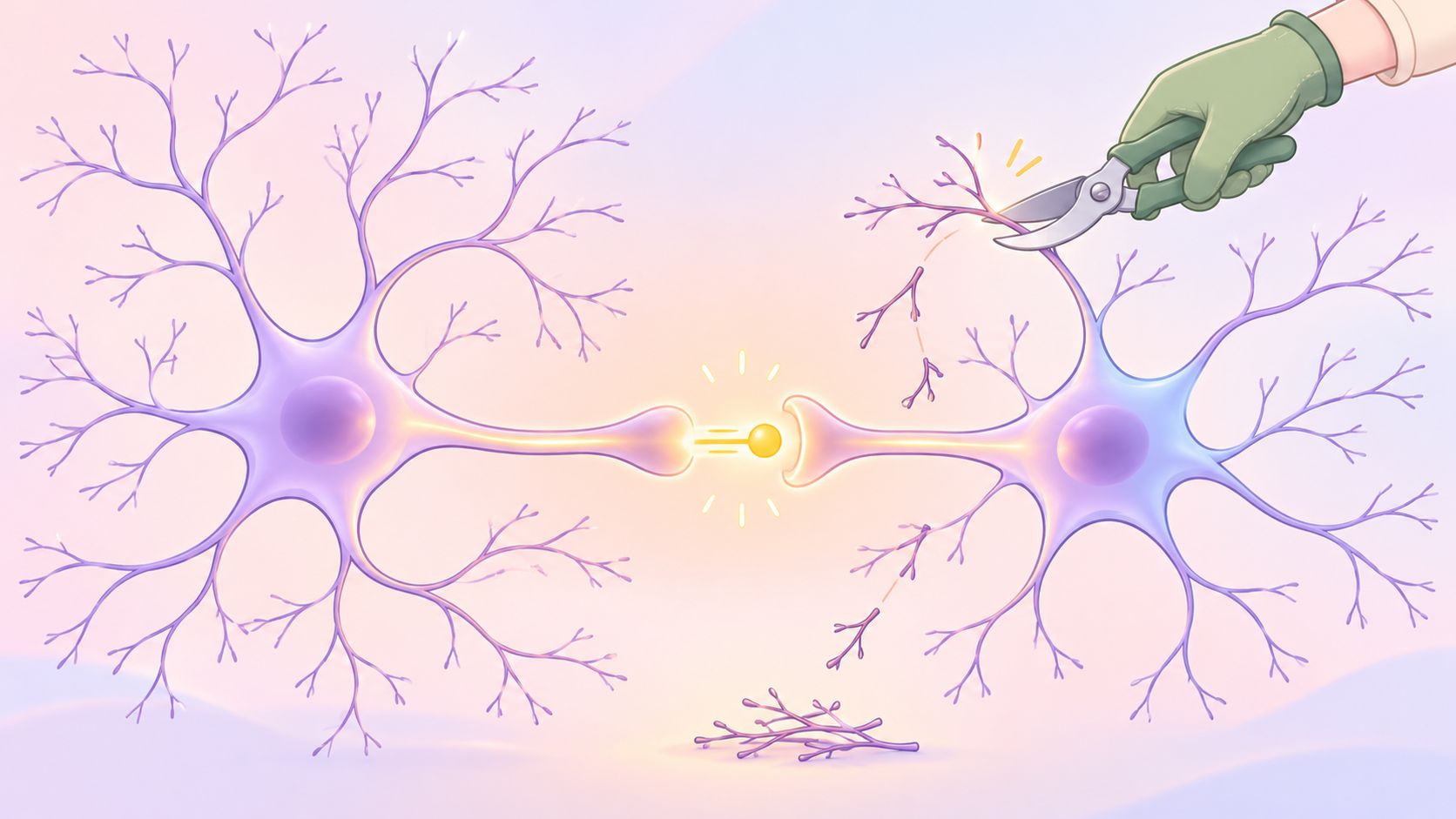
A synapse is a junction where a neuron communicates with another cell. Development involves both building connections and selectively removing some of them, rather than adding connections until the brain reaches its final form.
The cells and signals behind removal
Different components contribute to this process. Keeping their jobs separate prevents a common misunderstanding: synaptic pruning isn't one switch that can be turned up or down.
- Dendritic spines are tiny projections that receive many excitatory inputs. Scientists often count them as a proxy for those connections.
- Microglia are brain-resident immune cells that can engulf synaptic material. Engulfing material is one route involved in removing connections.
- Complement proteins, including C3, can participate in signals that help identify material for removal.
- Autophagy is a cell's internal recycling process. It isn't identical to a microglial cell swallowing material from outside itself.
- mTOR helps regulate cellular growth and metabolism. Strong activity in this pathway can suppress aspects of autophagy.
These processes interact, but a difference in one doesn't automatically tell us what all the others are doing. A protein measurement, a spine count, and an observation of microglial engulfment answer related but distinct questions.
Timing matters as much as quantity
Developmental pruning unfolds while circuits are still forming and changing. A snapshot from one stage can't establish what happened earlier or predict exactly what will happen later.
The proposed functional concern is signal-to-noise processing: whether a network can distinguish useful input from competing activity. Insufficient elimination could interfere with that distinction, while excessive or poorly timed removal could discard connections that support learning and flexibility.
Practical rule: More synapses doesn't automatically mean a better brain, and fewer synapses doesn't automatically mean a more efficient one.
Microglia also clear debris, release immune-signaling molecules called cytokines, and interact with cells involved in myelin formation. When a headline describes altered microglia, pruning may be only part of the biological story.
Too Little Pruning and Too Much Pruning
The familiar public explanation is straightforward: autistic brains don't remove enough connections, so extra connections remain. That interpretation fits one line of human tissue evidence, but it doesn't fit every experimental system.
In particular, changing a gene that affects neuronal excitability can produce a different result. Direction, genetic context, and developmental stage belong beside every pruning claim.
The review of immune-mediated pruning mechanisms describes 2023 work using cerebral organoids containing microglia and an autism-associated protein-truncating mutation in SCN2A. In that model, microglia eliminated more postsynaptic structures, a pattern described as over-pruning, rather than insufficient pruning.
Two pruning directions and the evidence behind each
| Pattern | Context where it appears | Proposed effect | Strongest evidence now |
|---|---|---|---|
| Insufficient pruning | Some postmortem cortex across childhood and adolescence | More spines remain, potentially changing signal-to-noise processing | Small, region-specific human tissue association |
| Excess pruning | SCN2A deficiency in mice and human cerebral organoids | Greater removal of postsynaptic structures at selected stages | Animal and organoid models, not a living-person test |
| Activity-dependent change | Inflammatory microglia studied under lower and higher synaptic activity | Lower activity impaired pruning; increased activity partly shifted the effect | Mechanistic model, not an autism outcome trial |
| Mixed or indirect | Immune-synaptic pathways across genetic contexts | Pruning may contribute in particular subsets | Reviews integrating several evidence types, with human causation unestablished |
A gene-specific result isn't an autism-wide result
SCN2A contributes to how readily neurons generate electrical signals. When scientists study a disruptive change in that gene, they're investigating a defined biological situation, not recreating every pathway that can contribute to autism.
Complementary mouse experiments connected SCN2A deficiency with partial microglial activation and C3-related engulfment at selected developmental stages. That supports a possible mechanism within the model, but it doesn't mean an autistic child's microglia are currently removing too many connections.
Nor do the opposing findings necessarily cancel each other out. Researchers could observe insufficient removal in one region or developmental context and excess removal in another without either observation being mistaken.
Activity can change the experimental result
Inflammation adds another complication. In a mechanistic model that combined inflammatory microglia with changing neural activity, pruning was impaired under reduced activity, while increased activity partly altered that effect.
That finding doesn't prescribe more stimulation, a particular learning program, or an anti-inflammatory treatment. It illustrates why describing microglia as overactive or underactive can conceal what investigators measured.
The strongest evidence for excessive pruning currently comes from animals, genetically defined cells, and organoids. Human autism evidence more often involves genetic associations, postmortem observations, or indirect imaging measures.
An observed difference could be a primary contributor, a response to altered neuronal activity, or an experimental marker of broader immune-neuronal change. A direct causal chain in people hasn't been established, and choosing among those explanations requires more than demonstrating that cells engulf synaptic material differently.
Three Research Models and What They Can Show
Suppose a parent encounters three headlines: extra connections in donated brains, excessive pruning in an organoid, and an autism-risk gene that changes immune-cell uptake. The headlines may sound contradictory because each compresses a different kind of experiment into the same word, pruning.
A better reading starts with the specimen. What scientists can conclude depends on whether they examined human tissue, manipulated laboratory-grown cells, or studied an animal.
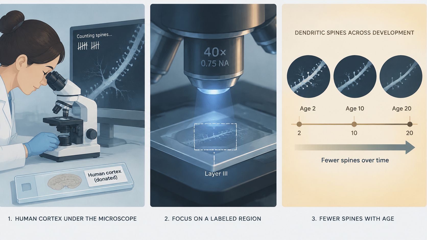
Donated human cortex
Postmortem tissue lets researchers inspect structures they can't routinely count during a clinic visit. Its strength is direct access to human brain material; its limitation is that it captures tissue after death rather than following development as it happens.
Allowed conclusion: The sampled groups differed in the structures remaining in the examined region. Unsupported conclusion: This count explains why a particular child shuts down in a classroom.
A tissue association cannot connect an individual's microscopic anatomy to a specific experience without additional evidence. It also isn't a measurement families can repeat at a checkup.
Organoids and complementary mouse experiments
Organoids are laboratory-grown tissue models, not miniature people. Researchers can introduce a chosen genetic change and examine interactions among human-derived cells under controlled conditions.
The Molecular Psychiatry paper by Wu and colleagues, published in 2024, investigated microglial over-pruning in SCN2A-deficient mice and human cerebral organoids. Pairing these systems can strengthen a mechanistic explanation without establishing that a corresponding treatment will help people.
Allowed conclusion: This genetic manipulation can alter pruning in these experimental systems. Unsupported conclusion: An intervention that changes the model should be given to a child to remove or preserve synapses.
Human microglia-neuron screens
A 2025 CRISPR-interference screen, described in the Molecular Psychiatry article on microglial endocytosis and pruning, identified ADNP as a modifier of these processes in human stem-cell-derived microglia and neurons. The mechanism by which microglia take up synaptic material remained incompletely defined.
Allowed conclusion: Changing a gene's activity can modify uptake in this cell system. Unsupported conclusion: Knowing that gene's status identifies a person's pruning problem or tells clinicians how to treat it.
For proximity to human brain anatomy, distinguish human tissue, then human cell models, then animals, then computational models. That ordering isn't a universal quality score, and none of these categories is equivalent to a human treatment trial.
Explanations of experience operate at another level. For example, monotropism and autistic attention concern patterns of attention, not a way to infer microscopic pruning from everyday behavior.
What This Means for Diagnosis and Daily Care
A developmental assessment can evaluate autism; it can't measure a person's synaptic-pruning status. No validated clinical test currently establishes that pruning caused someone's communication, sensory, or executive-function differences.
Autism assessment instead considers developmental history and observed patterns of social communication and restricted or repetitive behavior, including sensory features. A persuasive biological explanation isn't a substitute for understanding how someone developed and what they need now.
Keep peripheral immune findings in their proper place
A 2025 finding of impaired synaptosome phagocytosis in macrophages from autistic individuals, discussed in the Molecular Psychiatry publication on immune and synaptic processes, offered a possible peripheral immune clue. It did not directly measure synapse removal in the living brain.
Macrophages outside the brain and brain-resident microglia aren't interchangeable readouts. Finding that sampled cells take up isolated synaptic material differently doesn't establish what a person's brain cells are doing during daily life.
The same boundary applies to prognosis. A group-level pruning pattern doesn't determine an individual's intelligence, capacity to learn, future independence, or degree of support needed.
Choose care by the problem that needs help
Sleep disruption, infection, stress, and sensory overload deserve attention for their own effects. There isn't a validated clinical method for translating those concerns into a personalized instruction to increase or decrease pruning.
For a child distressed by a noisy lunchroom, useful questions concern sound intensity, recovery time, communication, and available accommodations. Calling the distress a pruning problem adds an unmeasured explanation without identifying the next practical step.
Understanding sensory processing differences can help families describe what happens, what triggers difficulty, and what support is workable. That description is more clinically useful than guessing how many connections remain.
Support communication, sensory needs, executive function, mental health, and access to school or work. Those are care goals in their own right, not indirect attempts to manipulate synapses.
Proposed targets such as mTOR, autophagy, complement, TREM2, and ADNP remain research directions rather than established disease-modifying autism treatments. A drug's use for another condition doesn't make it a safe or effective pruning intervention.
There's also a broader biological question: could an apparent pruning-related finding reflect altered immune signaling or myelin development instead? Because microglia participate in several neural processes, assigning a behavior to synapse removal alone can be premature.
Claims About Repairing Pruning That Do Not Hold
Autistic brains always have extra synapses is the claim to challenge first. It turns a conditional finding into a rule that opposing experimental patterns don't support.
Families shouldn't have to decode a sales pitch while learning developmental neuroscience. A useful credibility check is whether a claim acknowledges uncertainty or jumps directly from a biological possibility to something a family should buy or do.
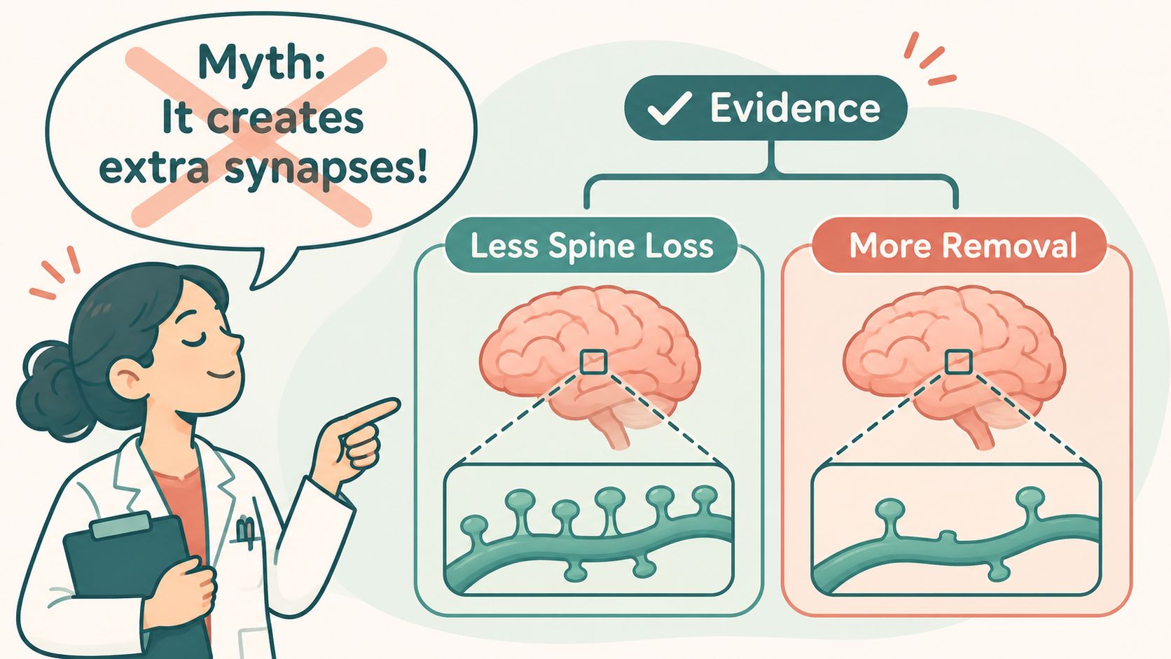
Common claims and their evidence mismatch
- Every autistic brain retains too many connections. Findings from selected human cortex don't establish a whole-brain rule, while genetically defined models can show greater removal instead.
- More synapses explain either exceptional intelligence or disability. Spine counts aren't intelligence scores, and a structural count alone can't identify the connections supporting a particular strength or difficulty.
- Sensory overload, speech delay, or shutdown reveals a pruning problem that a diet, supplement, or immune cleanse can repair. These experiences don't identify a pruning direction, and the evidence doesn't justify those interventions as ways to treat autism through synapse removal.
- A brain-type quiz or densely connected brain image identifies a pruning subtype. Neither a questionnaire nor a striking research illustration is a validated assay of an individual's synaptic elimination.
- If pruning is involved, autism means a damaged brain that needs cleanup. A proposed developmental mechanism doesn't establish that value judgment or erase the person's strengths, preferences, and support needs.
What a credible treatment claim would need
A convincing intervention claim would need more than a change in a laboratory marker. Researchers would need evidence that the intervention improves meaningful outcomes in people, with acceptable risks, rather than merely changing uptake or spine density in a model.
Be especially cautious when a seller treats opposite outcomes as equally persuasive. If both increasing and decreasing pruning are advertised as the same solution without identifying a validated patient group, the proposed mechanism isn't guiding care.
The credible stance is specific: pruning is interesting biology whose meaning is conditional on region, developmental stage, cell type, and gene. It isn't a lever families should try to pull themselves.
Choose an Evaluation When Daily Life Needs Clarity
If communication, sensory load, routines, school, work, or burnout are causing difficulty, the useful next step is an assessment of those experiences. Searching for a pruning score won't answer whether autism, ADHD, both, or another condition best explains the pattern.
A meaningful evaluation brings early history together with current demands. It should examine not only visible difficulties but also the effort required to compensate, rehearse interactions, or conceal distress.
Look for an explanation that leads to support
Masking can make a brief impression misleading. Women, girls, and people of color may need an assessment that pays close attention to experiences that aren't obvious to an observer.
At Sachs Center, we specialize in ADHD and autism, and our clinicians are trained to distinguish neurodivergence from CPTSD, generalized anxiety, depression, OCD, and personality disorders, including when several occur together. We use the ADOS as part of our autism diagnostic battery, alongside clinical assessment, rather than treating a synapse theory as diagnostic evidence.
Our comprehensive diagnostic assessment is conducted by secure video with experienced clinical psychologists for children, teens, and adults, including people whose experiences may be hidden by masking. Adult autism evaluation is built to recognize high-masking presentations, and a combined autism and ADHD report can recommend school or work accommodations when appropriate.
The useful outcome is a clearer account of your needs and a practical support plan, not a promise to measure or repair your synapses.
Bring concrete examples of situations that are difficult, what helps, and what remains exhausting even when you appear to manage. School records or recollections of early development can also help connect present concerns with a longer history.
After diagnostic clarity, therapy or coaching can focus on daily challenges without pretending to be a biological fix. Skip any provider who promises to diagnose a pruning subtype or tune synaptic elimination as routine autism care.
Sachs Center offers secure-video autism, ADHD, and combined evaluations for children, teens, and adults, with attention to presentations that masking can hide. If you want clarity about lived difficulties rather than an unmeasurable pruning theory, visit Sachs Center to book an evaluation and discuss a practical support plan.